
The iTrace vs. The NIDEK/Marco OPD III
HOW WE COMPARE
RAY TRACING
VS
DYNAMIC SKIASCOPY
The iTrace’s diagnostic aberrometry uses proprietary ocular Ray Tracing technology, which sends 256 consecutive beams of light into the eye one at a time and measures where each lands on the retina. The resulting pattern is measured to record aberrations, producing an exact simulation of how light functions when it enters the eye during the process of vision. This is the most accurate replication of ocular function, and the most accurate objective refraction possible.
The OPD III uses dynamic skiascopy, which sends a slit of light beams into the eye and measures aberrations as a function of the beams that bounce back out of the eye. Dynamic skiascopy has a “fatal flaw” — the fact that it has to interpolate results around a 2 mm blind spot in the center of the measurement field. [1] In cataract patients with smaller-than-average pupils, the central 2 mm is essential and guessing it makes overall data unreliable and difficult to use as a basis for critical decisions about patient care.
RAY TRACING
VS
DYNAMIC SKIASCOPY
WHY IT MATTERS
In Practical Terms: The iTrace vs The OPD III
In addition to the core differences in the technology used to scan patients, the following are key differences in the functionality and outputs available with the iTrace and the OPD III
PRE-SURGERY
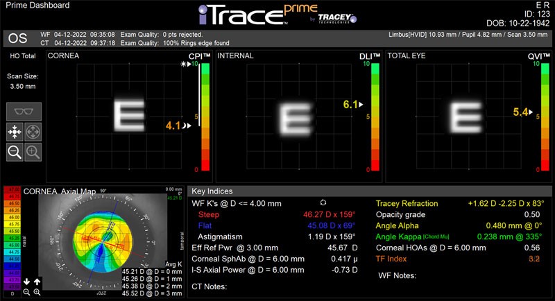
The iTrace Prime Dashboard
The iTrace Prime Dashboard includes these proprietary indices in a single display, making it simple to educate patients on the sources of their vision problems. It also gives cataract surgeons all the diagnostic information they need in a single display!
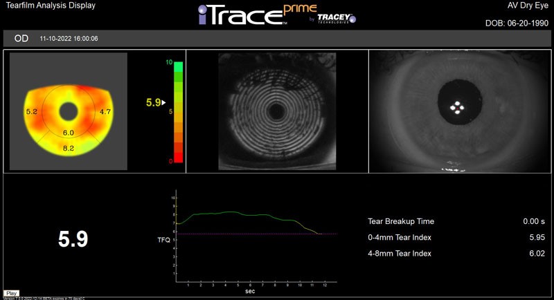
The Proprietary Tear Film Index
The OPD III doesn’t provide a similar way to assess a patient’s tear film quality, making it more difficult to ensure the best outcomes for confirmed and suspected dry eye patients.
POST-SURGERY
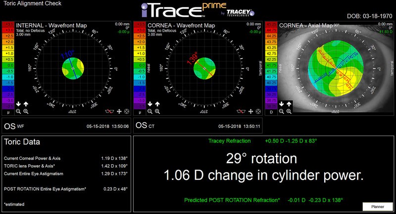
The iTrace Toric Check
You can use the iTrace to verify the placement of a toric IOL and determine if any additional rotation is needed to further improve vision. If you want to do a post-surgical confirmation of a toric IOL‘s placement with the OPD III, you’ll have to dilate the patient, and the results will be similar to an examination done with a slit lamp.
The iTrace generates a precise visual depiction of a toric IOL placement compared to the optimal axis location. It provides the patient’s current refraction and the exact degree of rotation it will take to get to the best outcome for a patient, guaranteeing the best possible results from toric lens surgery. All of this can be done without ever dilating a patient’s eyes!
Ready to Learn More
About the iTrace Advantage?
More importantly, when you purchase an iTrace, you’re getting the benefit of a team dedicated to customer support. At Tracey Technologies, our team is dedicated to your success using the iTrace — and we’ll make sure that you have the resources you need to better serve your patients and grow your practice!
To learn more about using the iTrace in your practice, schedule a demo, call or email us today!