Better Information, Better Outcomes

The iTrace Visual Analyzer is a first-of-its-kind tool that equips doctors to make the best decisions about correcting their patients’ vision.

iTrace Image

How the iTrace Benefits Doctors

What if you could install a single tool in your practice and get a complete, clear look at exactly how a patient sees the world? That’s what the iTrace was designed to do.

It’s a 5-in-1 tool that you can use in an exam and as part of the process of cataract surgery to make sure the decisions you make in your practice are in line with your patients’ best interests. Essentially, the iTrace takes all the guesswork out of visual analysis.

iTrace Image
iTrace Image

How the iTrace Benefits Patients

When patients see that their doctors are using the latest technology the industry has to offer, they’ll be more comfortable. They’ll notice that you’re taking the time to truly understand how they’re seeing the world around them, and they’ll appreciate the level of personal care they receive.

And, when you use the information provided by the iTrace, they’ll be thrilled with the results of their vision correction.

Improving the Patient Experience

Almost everything you do in your practice is done to provide patients with the best outcomes possible. The iTrace was developed to support doctors with diagnosis and surgical correction of vision, delivering the tools they need to create a superior patient experience.

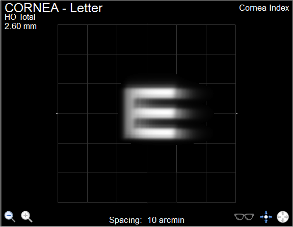
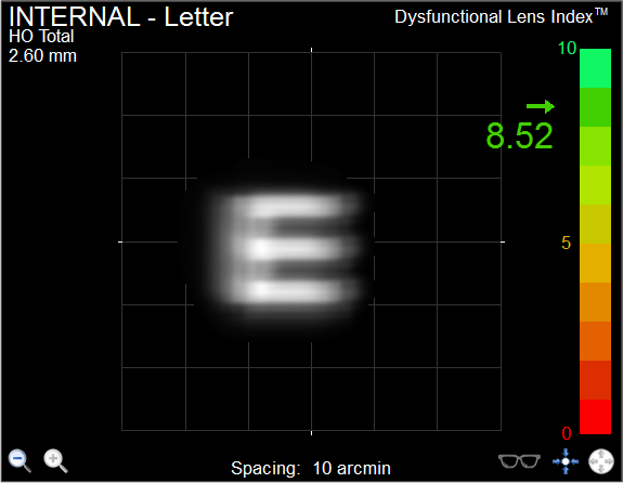
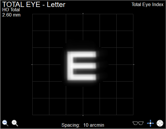
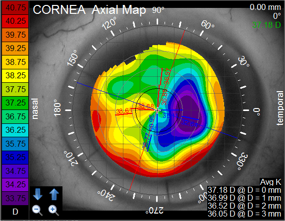
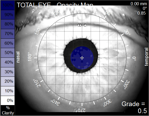
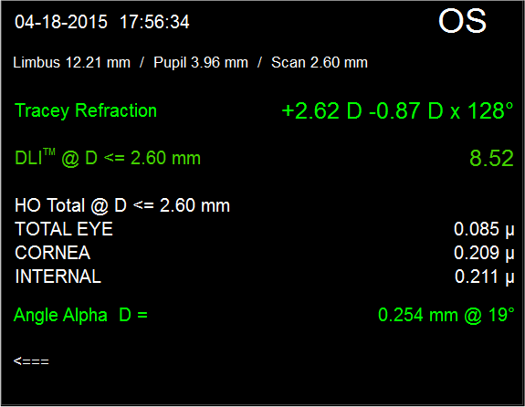
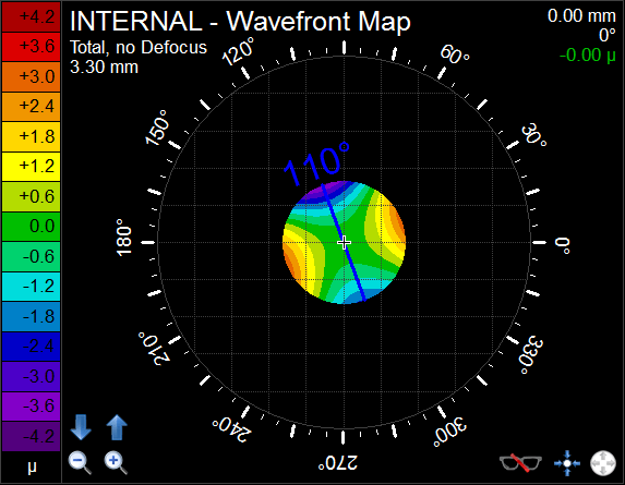
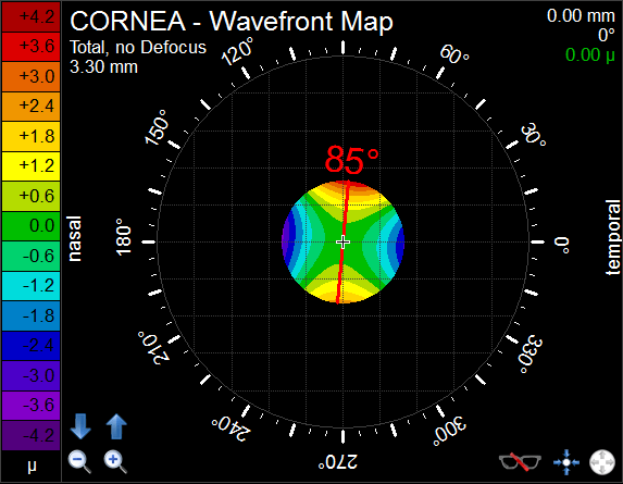
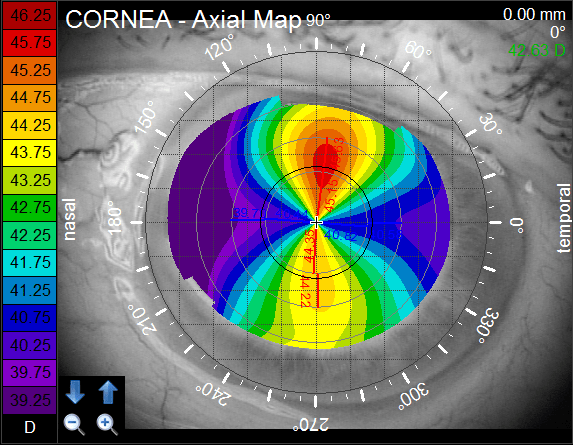
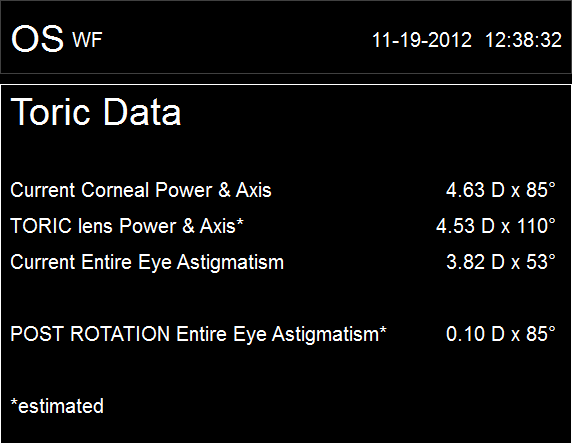
Using a series of calculations, the iTrace clearly illustrates how a patient’s vision quality is broken down between the cornea and the lens. This enables you to track vision loss over time and make an informed decision when surgical correction is necessary.

A simulated representation of the patient’s subjective corneal vision, isolated from the lens. This shows how well a patient would see if they had perfect lenticular vision.

A simulated representation of the patient’s subjective lenticular vision, isolated from the cornea. This shows how well a patient would see if they had perfect corneal vision.

A simulation of the patient’s total vision, giving a full representation of the patient’s vision from all components together. A patient can look at this screen and confirm their actual visual quality.

A corneal topography map shows the extent of patient’s astigmatism and other imperfections in the cornea.

An objective opacity map measuring obstructions (like a cataract) in the eye that may be contributing to visual issues for the patient.

Summary screen that specifies the eye being measured, the actual refraction data, and other metrics acquired from the one iTrace exam. Of particular interest is the objective measurement of the corneal imperfections characterized as HOAs. The corneal HOAs are an indicator of the cornea’s ability to support premium lens technologies.
The iTrace allows you to show patients a direct simulation of their vision after cataract surgery — with or without a toric IOL. With patients able to visualize the exact value of a toric IOL, they’ll upgrade to a toric lens more often.


The iTrace’s Toric Planner enables you to create a map of a patient’s eye and guide the precise placement of a toric lens during surgery. The Toric Check feature is used after surgery to compare the actual placement axis of the lens to the corneal steep axis and detect any changes in rotation necessary to achieve the optimal placement.

Current toric lens axis location.

Corneal steep axis and recommended toric placement.

Corneal Topography Map displaying the corneal curvature measured with the iTrace.

Comparison data on the power and axis of the cornea and of the toric lens with pre-rotation and post-rotation entire eye astigmatism.

Pre-rotation refraction, post-rotation refraction and precise rotation needed for the best possible vision correction so you can evaluate the potential improvement that can be achieved if a rotation is necessary.

TESTIMONIALS

SEE IF THE ITRACE IS RIGHT FOR YOU!